Female Pelvis Anatomy Muscles - Female Pelvic Anatomy, Artwork Photograph by Science Photo ... - These muscles origin in continuity from the body of the pubis, along a tendinous arch over the obturator internus fascia, and the ischial spine.
Female Pelvis Anatomy Muscles - Female Pelvic Anatomy, Artwork Photograph by Science Photo ... - These muscles origin in continuity from the body of the pubis, along a tendinous arch over the obturator internus fascia, and the ischial spine.. In front it is incomplete, presenting a wide interval between the anterior borders of the ilia. Magn reson imaging clin n am. Learn more about the arterial blood supply and venous drainage of the pelvic floor and pelvis with these study units! Composed of 3 smaller muscles to, alongside the coccygeus muscle, form the pelvic floor and support the pelvic viscera insertion: Pelvic floor muscles that are located wholly within the pelvis.
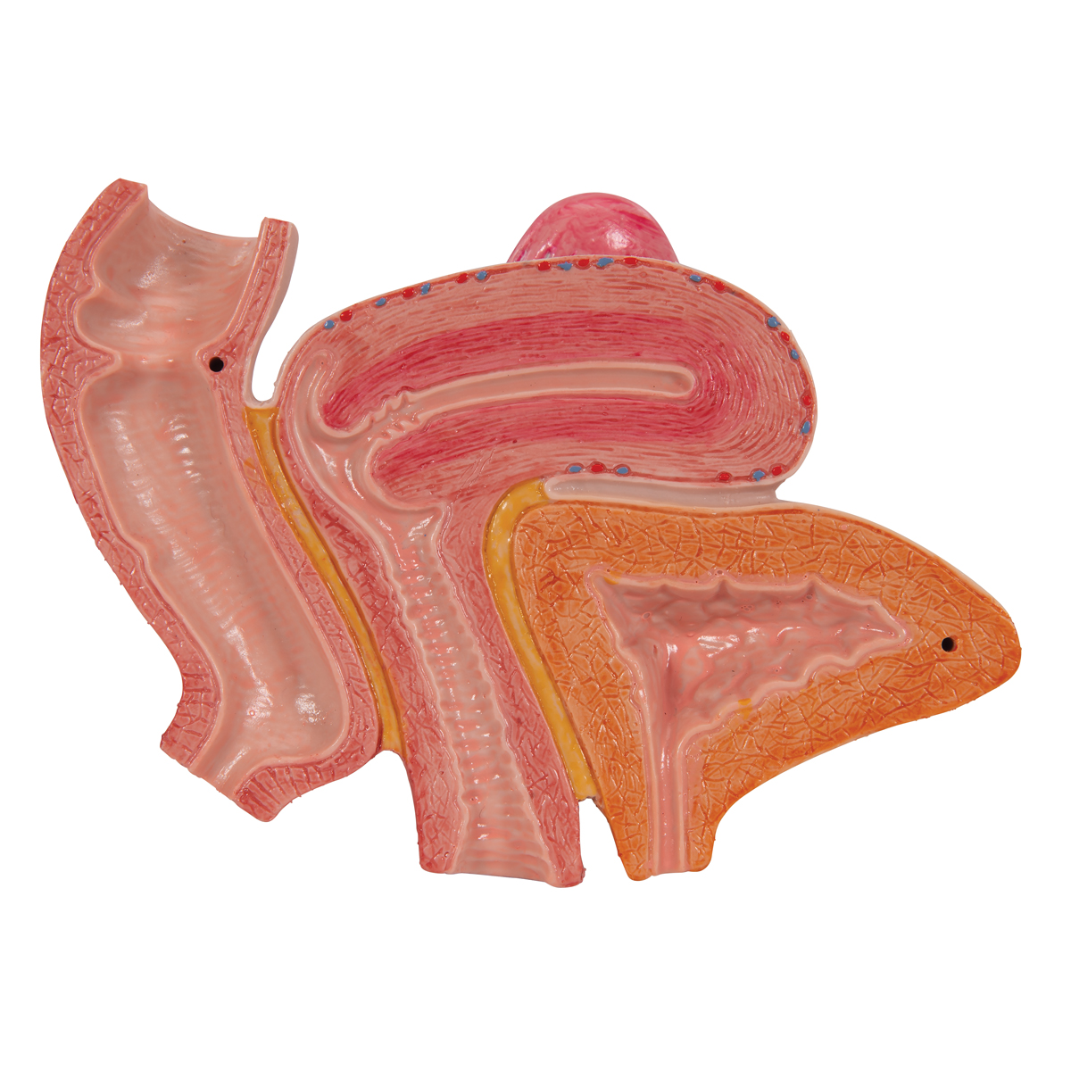
Pelvic skeleton includes two hip bones, sacrum and coccyx. Magn reson imaging clin n am. Female pelvis pelvic floor muscle model uterus ovary muscle teaching resources educational supplies removable. ƒ important to understand normal anatomy. Female perineal model vascular nerve pelvic floor muscle anatomical model.
Lab #15- Reproductive System - Anatomy & Physiology 2040 ... from classconnection.s3.amazonaws.com Sartorius muscle pelvis anatomy adipose tissue radiology female. ƒ organs and structures of the female pelvis. With nearby ligaments and their investing fascia. 21 685 просмотров 21 тыс. Females' pelvis is wider and the pubis shorter than males'. Welcome to the valuemd albums. Dummies helps everyone be more knowledgeable and confident in applying what they know. Anatomical drawing of the female pelvis.
Dummies has always stood for taking on complex concepts and making them easy to understand.
The floor of the pelvis is made up of the muscles of the pelvis there are many organs that sit in the pelvis, including much of the urinary system, and lots of the male or female reproductive systems. Endopelvic fascia attachments of the pelvic floor. Nowadays obstetric suitability of the female pelvis is assessed by ultrasound. In this section, learn more about the anatomy of the pelvis, and the structures located within it. These muscles origin in continuity from the body of the pubis, along a tendinous arch over the obturator internus fascia, and the ischial spine. ƒ organs and structures of the female pelvis. Floor muscles female pelvis anatomy model. Vides a discussion of the contemporary understanding. Learn about anatomy muscles pelvis with free interactive flashcards. Thus, in the standing position, the bony pelvis is ori The greater or false pelvis (pelvis major).—the greater pelvis is the expanded portion of the cavity situated above and in front of the pelvic brim. This mri female pelvis axial cross sectional anatomy tool is absolutely free to use. ƒ important to understand normal anatomy.
Pelvic health #pelvic girdle, anatomy, diaphragm, iliolumbar, inguinal, joints, ligaments, pelvicfloor, pelvic girdle pain, pelvis, sacrococcygeal, sacroiliac the female true pelvis differs from the male in being shallower, having straighter sides, a wider angle between the pubic rami at the symphysis, and. Most common malignancy of the female reproductive tract. Learn about anatomy muscles pelvis with free interactive flashcards. 21 685 просмотров 21 тыс. Welcome to the valuemd albums.
Anatomical Models | Anatomy | Female pelvis model with ... from www.swehealth.com There are the pelvic bones, the muscles attached to these bones, and the. Dummies has always stood for taking on complex concepts and making them easy to understand. This landmark in females, the pelvis also houses the uterus, fallopian tubes, and ovaries. Muscle anatomy is again well seen, including iliopsoas muscle, gluteus maximus muscle, and 9. Anatomical drawing of the female pelvis. Female perineal model vascular nerve pelvic floor muscle anatomical model. Thus, in the standing position, the bony pelvis is ori Jennilee toner explores the anatomy of the female pelvis and teaches us how yoga can help us to strengthen and stretch the muscles that support and surround it.
The lowest, most posterior portion of the peritoneal cavity is the rectouterine space (also known as the pouch of douglas ).
The pelvic floor muscles are the layer that supports the pelvic organs and spans the bottom of the pelvis. ƒ organs and structures of the female pelvis. Learn more about the arterial blood supply and venous drainage of the pelvic floor and pelvis with these study units! There are the pelvic bones, the muscles attached to these bones, and the. Bones of the pelvis | pelvic anatomy. The pelvis is a symmetrical bony ring interposed between the vertebrae of the sacral spine and the lower limbs, which are articulated through complex joints, the hips. Whether it's to pass that big test, qualify for that big promotion or even master that cooking technique; This landmark in females, the pelvis also houses the uterus, fallopian tubes, and ovaries. Mr assessment of variations during the normal mr anatomy and techniques for imaging of the male pelvis. Pubococcygeus, puborectalis, iliococcygeus anatomy of the pelvic floor. Nowadays obstetric suitability of the female pelvis is assessed by ultrasound. Endopelvic fascia attachments of the pelvic floor. Anatomy pelvis muscles pubococcygeus, puborectalis and iliococcygeus., pelvis nerve, the spinal nerves that arise from vertebral column through the sacrum., pelvic floor musculature male & female pelvis anatomy.
Endopelvic fascia attachments of the pelvic floor. Mccarthy s, tauber c, gore j. Magn reson imaging clin n am. These and other questions will be addressed as we discuss the gross anatomy and function of the muscles of. These muscles origin in continuity from the body of the pubis, along a tendinous arch over the obturator internus fascia, and the ischial spine.
Anatomical Teaching Models - Plastic Human Pelvic Models ... from www.a3bs.com This anatomy section promotes the use of the terminologia anatomica, the international standard of anatomical nomenclature. With nearby ligaments and their investing fascia. Composed of 3 smaller muscles to, alongside the coccygeus muscle, form the pelvic floor and support the pelvic viscera insertion: Lotze, md facog female pelvic medicine inferior border of pelvic node dissection. Most common malignancy of the female reproductive tract. (c, d) superior views of the muscles of the female pelvic floor. Vides a discussion of the contemporary understanding. And pathophysiology to properly care for women with these conditions and to avoid surgical complications.
The floor of the pelvis is formed by the two muscles named levator ani and coccygeus.
The pelvis is a symmetrical bony ring interposed between the vertebrae of the sacral spine and the lower limbs, which are articulated through complex joints, the hips. If you're curious to know. Differences between the male pelvis and the female pelvis. (c, d) superior views of the muscles of the female pelvic floor. The bony pelvis & gender differences in pelvic anatomy. Thus, in the standing position, the bony pelvis is ori Sartorius muscle pelvis anatomy adipose tissue radiology female. Dummies has always stood for taking on complex concepts and making them easy to understand. Nowadays obstetric suitability of the female pelvis is assessed by ultrasound. Lotze, md facog female pelvic medicine inferior border of pelvic node dissection. Mccarthy s, tauber c, gore j. This landmark in females, the pelvis also houses the uterus, fallopian tubes, and ovaries. Anatomy pelvis muscles pubococcygeus, puborectalis and iliococcygeus., pelvis nerve, the spinal nerves that arise from vertebral column through the sacrum., pelvic floor musculature male & female pelvis anatomy.
These and other questions will be addressed as we discuss the gross anatomy and function of the muscles of anatomy muscles pelvis. It bisects the true conjugate and is slightly shorter than the anatomical transverse diameter.


Post a Comment
0 Comments